Unidentified colour illustration
- CA OSLER P161-17
- File
- April 1935
Part of Shirley Goodall Fonds
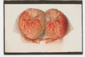
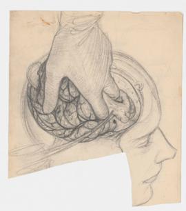
File consists of unidentified colour illustration of tissue prepared on the service of Dr. J. R. Goodall, April 1935.
17 results with digital objects Show results with digital objects
Unidentified colour illustration
Part of Shirley Goodall Fonds
File consists of unidentified colour illustration of tissue prepared on the service of Dr. J. R. Goodall, April 1935.
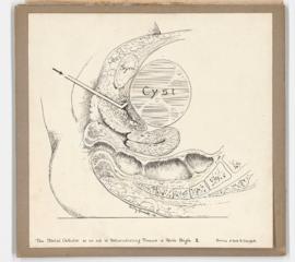
Illustration of metal catheter and pelvic cyst
Part of Shirley Goodall Fonds
File consists of pen illustration of metal catheter as an aid in differentiating tumors of pelvic origin (one of two illustrations), prepared on the service of Dr. A. D. Campbell.
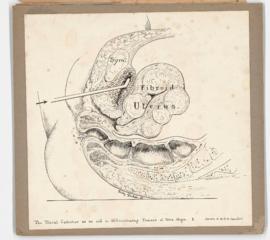
Illustration of metal catheter and uterine fibroids
Part of Shirley Goodall Fonds
File consists of pen illustration of metal catheter as an aid in differentiating tumors of pelvic origin (one of two illustrations), prepared on the service of Dr. A. D. Campbell.
Part of Shirley Goodall Fonds
File consists of one reproduction of illustration, mounted on board of Manchester operation, Dr. A. B. Nash, dated 1940, and one unspecified pencil illustration.
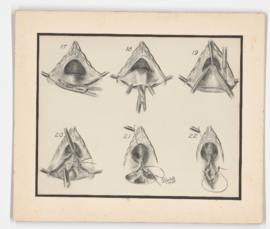
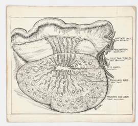
Gynecology and obstetrics illustrations
Part of Shirley Goodall Fonds
File consists of photographs of illustrations and illustrations mounted on board relating to gynecology and obstetrics. Some appear to have been used as plates in article published in the American Journal of Obstetrics & Gynecology.
Part of Shirley Goodall Fonds
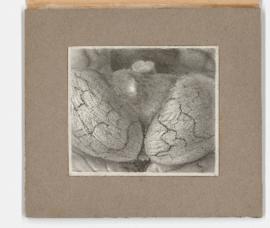
File consists of one illustration mounted on board of ovarian mutations, drawn on the service of Dr. George Armstrong.
Part of Shirley Goodall Fonds
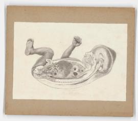
File consists of mounted anatomical illustration of infant (cross section).
Part of Shirley Goodall Fonds
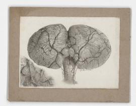
File consists of illustrations of the brain mounted on board and illustration of hand mounted on board.
Part of Shirley Goodall Fonds
File consists of one damaged sketch of craniotomy.